


Systemic lupus erythematosus (SLE) is a complex autoimmune disease characterized by the immune system attacking healthy tissues. The etiology of SLE remains unclear, hindering the development of personalized treatment strategies.
A recent study has identified a potential mechanism underlying SLE pathogenesis (Figure 1). The findings suggest that the transcription factor ETV5 plays a critical role in promoting the differentiation of T cells into T follicular helper (TFH) cells.
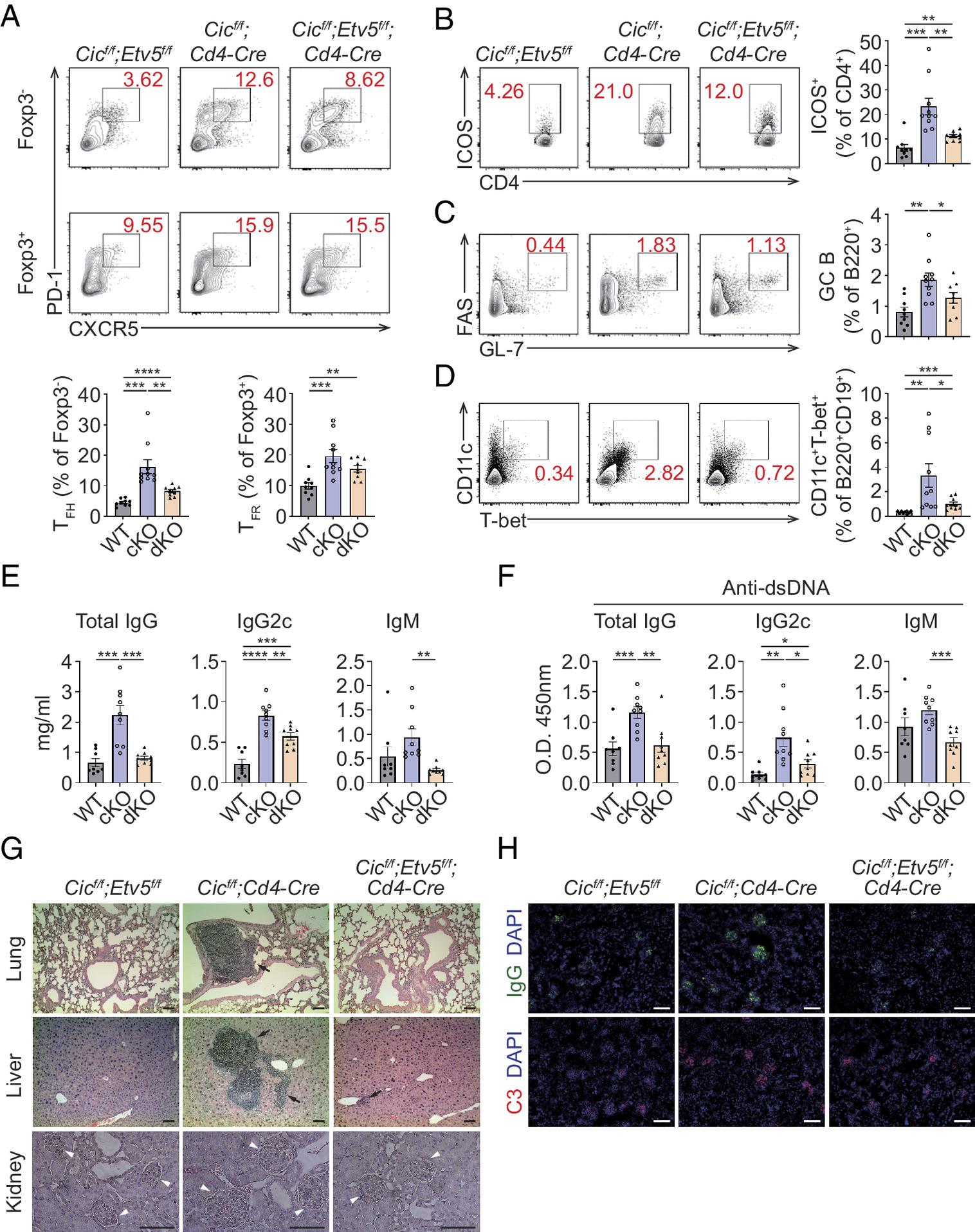
Figure 1: Recovery of autoimmune-like phenotypes in T cell–specific Cic and Etv5 double-null mice. (A–D) Flow cytometry of TFH and TFR cells (A), CD4+ICOS+ T cells (B), GC B cells (C), and CD11c+Tbet+ B cells (D) from spleens of 8-mo-old Cicf/f;Etv5f/f (n = 9), Cicf/f;Cd4-Cre Cre (n = 10), and Cicf/f;Etv5f/f;Cd4-Cre (n = 9) mice. Each dot in the graphs represents an individual mouse. These data represent the combination of results from three independent experiments. (E and F) Serum levels of total IgG, IgG2c, and IgM (E) and anti-dsDNA antibodies (F) in Cicf/f;Etv5f/f f (n = 8), Cicf/f;Cd4-Cre (n = 9), and Cicf/f;Etv5f/f;Cd4-Cre (n = 9) mice as measured by the ELISA. The graphs show the data as mean ± SEM. Each dot in the graphs represents an individual mouse. WT: Cicf/f;Etv5f/f, cKO: Cicf/f;Cd4-Cre, and dKO: Cicf/f;Etv5f/f;Cd4-Cre. *P < 0.05, **P < 0.01, ***P < 0.001, and ****P < 0.0001. (G) Hematoxylin and eosin staining of the liver, lung, and kidney of 8-mo-old Cicf/f;Etv5f/f, Cicf/f;Cd4-Cre, and Cicf/f;Etv5f/f;Cd4-Cre mice. Black arrows and white arrowheads indicate infiltrating immune cells and glomeruli, respectively. Two independent experiments were performed. (Scale bar, 100 μm.) (H) Representative immunofluorescence images of kidney sections of 8-mo-old Cicf/f;Etv5f/f, Cicf/f;Cd4-Cre, and Cicf/f;Etv5f/f;Cd4-Cre mice. Kidney sections were stained with anti-IgG (green), anti-C3 (red), and DAPI (blue). Two independent experiments were performed. (Scale bar, 100 μm.)
Normally, TFH cells play a beneficial role by assisting B cells in generating antibodies against pathogens. However, excessive TFH cell activity can lead to B cell hyperactivity and the production of autoantibodies, which target the body’s own tissues.
The study employed both in vivo and in vitro models to demonstrate the link between ETV5, TFH cell differentiation, and Lupus development. ETV5-deficient lupus mice exhibited reduced autoimmune symptoms, including lower autoantibody concentrations, decreased immune cell infiltration into tissues, and attenuated renal glomerulonephritis.
Furthermore, the research team identified osteopontin (OPN) as a downstream target of ETV5. ETV5 upregulates OPN expression, which in turn activates the AKT signaling pathway, ultimately promoting TFH cell differentiation.
Importantly, the findings translated from the animal models to human patients. ETV5 and OPN levels were significantly elevated in CD4+ T cells isolated from SLE patients compared to healthy controls. Additionally, ETV5 and OPN expression levels correlated with disease activity and autoantibody concentrations.
This research offers valuable insights into the mechanisms underlying SLE. Targeting the ETV5-OPN pathway holds promise for the development of novel therapeutic strategies for this debilitating autoimmune disease.
Journal article: Park, J., et al., 2024. ETV5 promotes lupus pathogenesis and follicular helper T cell differentiation by inducing osteopontin expression. Proceedings of the National Academy of Sciences.
Summary by Stefan Botha


