


Stroke is a leading cause of death and long-term disability, with ischemic strokes making up 85% of cases. Diagnosing stroke accurately and predicting disease progression remains challenging. In recent years, Immuno-MRI has emerged as a promising tool for detecting neuro-inflammation and predicting stroke outcomes. A 2024 review, published in Neuroscience, explores how immuno-MRI can revolutionize stroke diagnosis and treatment.
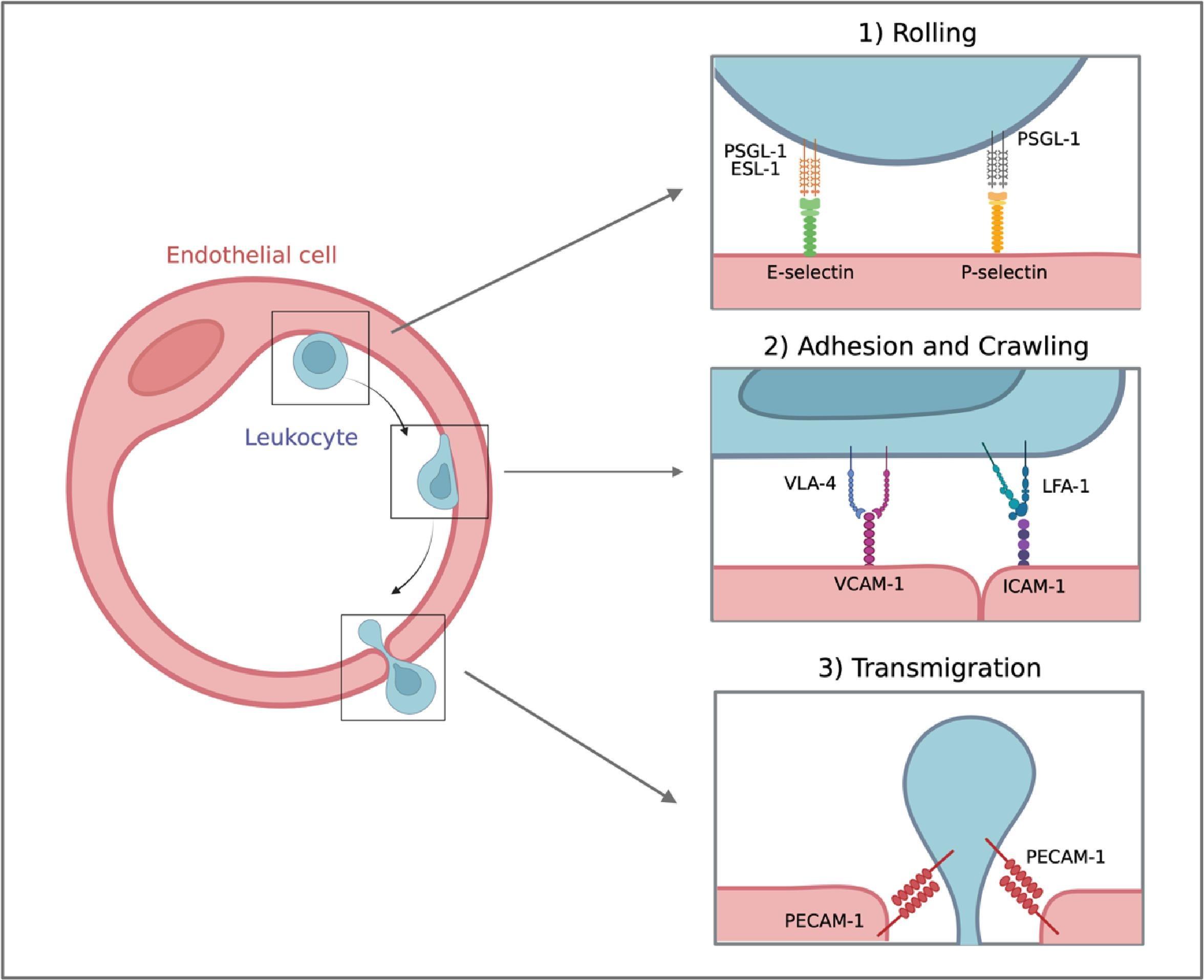
Figure 1: Immune cell infiltration across the BBB is a complex, multistep process involving the interaction between circulating leukocytes and adhesion molecules expressed on the surface of activated endothelial cells
Immuno-MRI Technology
Immuno-MRI uses iron oxide particles conjugated with antibodies to detect specific biomarkers, such as adhesion molecules, on the surface of activated endothelial cells. These molecules are crucial indicators of inflammation, especially during the acute phase of a stroke. Traditional imaging methods, like CT and standard MRI, do not provide information on inflammation at the cellular level. Immuno-MRI, however, can capture high-resolution images of inflammation in real-time without ionizing radiation.
Immuno-MRI is particularly useful for detecting stroke-induced neuro-inflammation, which begins with blood-brain barrier (BBB) permeability and immune cell infiltration. Preclinical studies demonstrated that targeting adhesion molecules like VCAM-1 and P-selectin with immuno-MRI allows early detection of endothelial activation during stroke. This ability to visualize inflammation at such an early stage could enable more personalized treatments, such as immune-modulating therapies, which have shown promise in reducing stroke damage.
Furthermore, immuno-MRI could differentiate between transient ischemic attacks (TIAs) and strokes, which is crucial in preventing future stroke events. In TIA models, immuno-MRI targeting P-selectin revealed endothelial activation even in the absence of acute infarction.
Clinical Implications
The ability to monitor stroke-induced inflammation non-invasively and in real-time holds great potential for clinical use. Immuno-MRI could improve patient outcomes by helping clinicians identify candidates for reperfusion therapies and monitor treatment efficacy. The next step for immuno-MRI is clinical trials to assess its applicability in routine stroke management. Immuno-MRI offers an innovative and non-invasive method to detect neuro-inflammation during stroke. Its ability to visualize cellular-level changes in the brain could revolutionize stroke diagnosis and prognosis, leading to more targeted therapies and improved patient outcomes.
Journal article: Fournier, Antoine Philippe, et al. “Immuno-MRI for Stroke Diagnosis and Prognosis.” Neuroscience, 21 Dec. 2023.
Summary by Faith Oluwamakinde


