In a ground-breaking study published in Viruses, researchers delve into the dual role of autophagy and apoptosis in the replication of the rabies virus (RABV). The intricate balance between these two cellular processes is vital to understanding the pathogenesis of rabies and developing future therapeutic approaches.

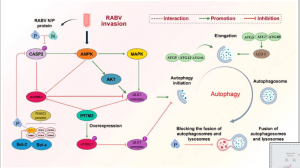
Figure 1: Rabies Invasion. Mechanism of RABV-induced incomplete autophagy. (1) RABV activates the initiation of autophagy. RABV activates the AMPK signaling pathway upon invasion. On the one hand, activated AMPK inhibits mTORC1, thus blocking the inhibition of mTORC1 on the ULK1 complex. On the other hand, AMPK can positively regulate two factors, AKT and MAPK, to activate the ULK1 complex and prompt the initiation of autophagy. In addition, the P protein of rabies virus binds to Beclin1 in the PI3KC3 complex and reduces the phosphorylation of CASP2, which not only positively regulates the AMPK signaling pathway but also negatively regulates the mTORC1 signaling pathway, which activates the initiation of autophagy. (2) RABV prevents the fusion of autophagosome and lysosome: the P protein of RABV binds to Beclin1, wraps immature autophagosomes, inhibits the fusion of autophagosome and lysosome, and blocks the degradation of autophagosomes. (3) IFIM3 was able to directly inhibit the ULK1 complex and promote mTORC1 phosphorylation, indirectly inhibiting the ULK1 complex. Blocked the initiation of autophagy induced by RABV invasion.
Autophagy, a cellular process that maintains homeostasis by degrading damaged cellular components, is hijacked by RABV to promote its replication. Instead of merely serving a protective role, autophagy facilitates the replication of the virus in host cells. On the other hand, apoptosis, which is the programmed cell death mechanism, is a defence response activated by host cells when they are infected. However, RABV has evolved mechanisms to inhibit apoptosis, allowing the virus more time to replicate and spread before the host cell is destroyed.
The study suggests that this interplay between autophagy and apoptosis is a major determinant of disease progression in rabies. Researchers observed that during early stages of infection, autophagy is upregulated to benefit the virus, while apoptosis is delayed to prolong cell survival. As the infection progresses, apoptosis eventually ensues, leading to the death of infected cells and contributing to the severe neurological damage seen in rabies.
The research also highlights potential therapeutic interventions. By targeting autophagy pathways or restoring apoptotic function in infected cells, it may be possible to reduce viral replication and limit the extent of brain damage in rabies patients. This study offers a fresh perspective on how cellular processes are manipulated by RABV, opening new avenues for therapeutic strategies against this deadly disease.
Journal article: Li, Saisai, et al. “Autophagy and Apoptosis in Rabies Virus Replication.” Cells.
Summary by Faith Oluwamakinde


