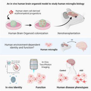
Microglia are special immune cells in the brain that are important for brain development and disease. However, studying them has been challenging because they can’t be easily studied outside the human brain. Recently, scientists have developed a model called an organoid, which is a small version of human brain tissue (Figure 1). This organoid model allows researchers to study microglia in a living human environment for the first time.
In a new study, researchers examined patient-derived microglia from children macrocephalic autism to understand how the brain environment influences their development and function. They found that certain proteins are crucial for microglia to function properly and that these proteins appear early in brain development.
In contrast to earlier models, the scientists developed a human brain organoid that contained microglia and simulated a human brain environment. This breakthrough enabled them to examine how the environment affects microglia during the development of the brain. They discovered that a specific protein called SALL1 appeared as early as eleven weeks into development, which confirmed the identity of microglia and supported their mature function. Additionally, they observed that certain factors specific to the brain environment, such as the proteins TMEM119 and P2RY12, were essential for the proper functioning of microglia.
This research enhances our understanding of diseases like autism and Alzheimer’s, which involve problems with brain development and function. It also highlights the significance of the brain environment in microglia’s functioning. However, it’s important to note that this study had a small sample size, and more research is required to confirm the findings. In the future, the researchers plan to study more microglia and explore their role in other diseases as well.
Journal article: Schafer., S.T., et al., 2023. An in vivo neuroimmune organoid model to study human microglia phenotypes. Cell.
Summary by Stefan Botha


