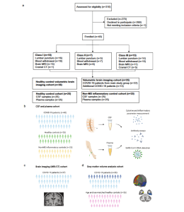
It has become known that during COVID-19 infection, individuals may lose their sense of taste and smell. In some, the disease might cause damage to the nervous system which may lead to concentration issues or even stroke. In a new study (Figure 1), researchers have described the effects of COVID-19 on the neurological system and the subsequent development of ‘neuro-COVID’.

Figure 1: CONSORT diagram and schemes illustrating the project design. a) Consort flow diagram. Patients who tested positive for SARS-CoV-2 were assessed for eligibility (n = 310), of which 269 declined to participate and 1 failed to meet inclusion criteria. Enrolled patients (n = 40) were allocated to different severity classes of Neuro-COVID according to Fotuhi et. al.13 with 18 in class I, 7 in class II and 15 in class III. Schemes illustrating the study design: b Paired cerebrospinal fluid (CSF) and plasma samples were collected from 40 COVID-19 patients. Paired CSF and plasma samples from healthy (n = 25) and non-MS inflammatory neurological disease controls (n = 25) were retrospectively obtained. c In 37 of the COVID-19 patients, a contrast-enhanced MRI or CT scan was conducted and evaluated by a board-certified neuroradiologist. d Brain volumetric analysis was performed in 35 COVID-19 patients. This cohort included 22 patients of the main study cohort from whom Magnetization prepared—rapid gradient echo (MPRAGE) pulse sequences and paired CSF and plasma samples were obtained (light blue) and an additional 13 patients who underwent brain MRI during COVID-19 infection (dark blue). A cohort of 36 healthy age and sex-matched individuals served as the control group. b–d Created with Biorender.com.
Through the analysis of cerebrospinal fluid and blood plasma from people suffering from COVID-19, the team were able to detect and predict different severities of neuro-COVID. This study may offer insights into how we can mitigate the effects of COVID-19 on the neurological system.
Studying 40 Covid-19 patients with neurological symptoms of varying disease, they measured the brain structures of these patients and and surveyed the participants 13 months after their illness to described lasting COVID-19 symptoms.
They were able to show affected individuals had impairment of the blood-brain barrier which was possibly triggered by the inflammatory “cytokine storm”.
They then were able to show how antibodies that targeted certain parts of the body, especially in the brain and cause damage i.e. an autoimmune response, as well as identifying how the brains’ microglia (immune cells) were activated during infection.
In order to develop a blood test to help diagnose and treat those with neuro-COVID, they found a few biomarkers which could point to potential targets for drugs subsequently mitigating the damage due to a Covid-19 infection. One of the biomarkers identified in blood, the factor MCP-3.
Journal article: Manina M. Etter, M.M, et al., 2022. Severe Neuro-COVID is associated with peripheral immune signatures, autoimmunity and neurodegeneration: a prospective cross-sectional study. Nature Communications.
Summary by Stefan Botha


